Spina Bifida
Spina bifida is a malformation caused by the poor development of the spine and spinal cord in the uterus, leaving an opening in the spine. Learn about the four different types of spina bifida, their causes, and how it is diagnosed.
Spina bifida means that the bones that protect the spinal cord are not fully formed.
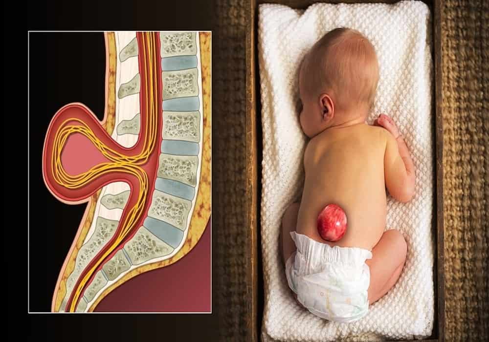
In babies with spina bifida, the cerebrospinal fluid (CSF), nerves, and the membrane around the spinal cord form a protrusion that protrudes from the baby’s back.
There are four common types of spina bifida: myelomeningocele, meningocele, lipomyelomeningocele, and spina bifida occulta.
The causes of this malformation are unknown, but spina bifida and other neural tube defects are less likely to occur when women receive enough folic acid.
What is spina bifida?
The spinal cord is a thick bundle of nerves that carries messages between the brain and the rest of the body. It floats in a fluid called cerebrospinal fluid (CSF). This fluid nourishes and protects the brain and spinal cord. The CSF is covered with a lining made of three thin layers called the meninges. This lining is normally protected by the bones of the back (the vertebrae).
In people with spina bifida, the bones that protect the baby’s spinal cord are not fully formed during pregnancy. The lining, CSF, and spinal cord are left unprotected, which occurs very early in the baby’s growth.
Spina bifida can affect any part of a baby’s back, between the head and the hips, but most often in the lower back. This region is called the lumbar or lumbosacral spine.
Children with spina bifida may have health problems related to the disorder, they may experience loss of sensation in their legs, have a reduced ability to move them, or even become paralyzed. They may also have problems with their bladder and bowel functions.
Types of spina bifida
There are four main types of spina bifida:
1. Spina bifida occulta
2. Lipomyelomeningocele
3. Meningocele
4 Myelomeningocele “Meningo” refers to the wall of the spinal canal. “Myelo” refers to the spinal cord itself. “Célé” means something bulging.
Spina bifida occulta
The occulta type is the less severe form of spina bifida. It occurs when a small section of the outer part of the vertebrae (the bones of the spine) has not completely closed, leaving an opening. In this type of spina bifida, the spinal cord and its layers (the meninges) are usually not damaged and do not protrude or overflow through the opening. There may be a dimple, hairy area, birthmark, or fatty lump in the affected area. This type of spina bifida may not be detected until birth. Many people can have this type of spina bifida and not know it.
Lipomyelomeningocele
A lipomyelomeningocele is a form of spina bifida where the outer part of the vertebrae does not fully enclose, exposing a gaping opening. Some abnormal fatty tissue is forced through the opening and can cause compression of the nerves.
Meningocele
A meningocele is a more severe form of spina bifida. It occurs when the outer part of the vertebrae has not been fully closed, leaving an opening. The spinal cord itself may not be affected, but its protective coverings (the meninges) can be damaged and forced through the opening to form a sac, often covered with skin, that contains CSF. In meningocele, the spinal cord is retained in the area of the back where it is normally found. This means that children expecting meningocele can have normal mobility and tenderness in their legs and feet.
Myelomeningocele
A myelomeningocele is the most severe form of spina bifida. It occurs when the outer part of the vertebrae does not fully close, creating an opening. In the case of myelomeningocele, the lining of the spinal cord (the meninges) and the spinal cord itself are forced through the opening. Usually, a liquid-filled sac covered with a thin membrane forms and can easily rupture, exposing its delicate contents.
Since part of the spinal cord overflows into the sac, the spinal cord does not develop properly and the nerves are damaged. Most children with myelomeningocele have motor and sensation problems in their legs and feet, and may be paralyzed.
Diagnosis of spina bifida
Spina bifida can be diagnosed during pregnancy or after the baby is born.
During pregnancy
Tests done during pregnancy can indicate that the baby has a high chance of having this malformation.
Alpha-fetoprotein (AFP) blood test – AFP is a protein produced by unborn babies. AFP crosses the placenta to the mother. A test measures the levels of AFP in the mother’s blood. If they are high, it may mean the baby has this malformation.
Ultrasound – This is a common test during pregnancy that allows healthcare professionals to see images of the unborn baby. In some cases, an ultrasound can show the baby has this malformation.
Amniocentesis – This is a small sample of amniotic fluid from the mother’s uterus. If this fluid has an above average AFP level, then the baby could have this malformation.
Fetal Magnetic Reasoning Imaging (MRI) – If initial tests suggest there is a high likelihood of this malformation, a fetal MRI may be done. The MRI performed in this case on the pregnant mother makes it possible to assess the unborn baby.
Spina bifida occulta may not be diagnosed until late childhood, adulthood, or not be diagnosed at all.
Treatment of spina bifida
The meningocele where only the meninges are pushed through the opening and the myelomeningocele where the meninges and spinal cord are forced through the opening are both treated with surgery. Older infants and young children with lipomyelomeningocele may require surgery if they develop symptoms. Spina bifida occulta usually does not require treatment.
The surgery can be done before your baby is born, before the 26th week of pregnancy, or within two to three days after birth.
Surgery before your baby is born
Sometimes when a baby is diagnosed with this malformation, especially myelomeningocele, surgery may be possible before the 26th week of pregnancy. It is performed by opening the mother’s uterus and repairing the defect in the baby’s spinal cord while it is still in the womb.
Some studies suggest that this type of intervention prevents further damage to the spinal cord and decreases the possibility of needing treatment for hydrocephalus. Your neurosurgeon and obstetrician will tell you if this option is possible for you and your baby.
Surgery after the birth of your baby
If your baby was diagnosed with this malformation during pregnancy, you may need to give birth in a high-risk pregnancy center. Your obstetrician will tell you if this is necessary. After childbirth, the sac cannot stay outside the body for a long time. It can tear or become infected, or it may already be ruptured, exposing its delicate contents. Your baby will have an operation to repair the sac within two to three days of birth.
A neurosurgeon will perform the operation. A neurosurgeon is a doctor who performs operations on the brain and spinal cord.
Your baby may need to have magnetic resonance imaging (MRI) before surgery to help surgeons see the spinal cord better. An MRI is a test that takes special pictures of the inside of the body. To have an MRI, the patient must remain still while the photos are taken. Some children may need sedatives to help them stay still during the test.
The operation aims two things:
- close the skin over the bag
- avoiding infections and other damage to the spinal cord
If your baby moves his legs and feet less or is unable to move them at all, the operation will not restore movement and sensitivity to the baby’s legs and feet.
Health problems associated with or caused by spina bifida
Every child with spina bifida is different with their own medical, mobility and learning challenges. Some children may only be mildly affected while others may have more severe disabilities. Being born with spina bifida brings challenges for life. Your child’s healthcare team will work with you to help your child reach their greatest potential.
The following health problems are common in children with spina bifida:
Hydrocephalus
About 80% of babies born with spina bifida, mainly those with myelomeningocele, will also have hydrocephalus. Hydrocephalus is characterized by the abnormal buildup of CSF in the ventricles inside the brain.
Chiari malformation
Almost all babies born with myelomeningocele have a type 2 Chiari malformation. This is when the lower part of the brain (the brainstem) sits too low in the upper area of your child’s spine. Some children with type 2 Chiari malformation may have feeding problems (eg, weak sucking when feeding, gagging, choking, difficulty swallowing), breathing problems, and some may have weakness in their stomach. arm level. Surgery may be needed to take the pressure off the lower part of the brain.
Physiological function of the legs (mobility) and sensation
In children with spina bifida, the nerves in the neural canal are often damaged or malformed; therefore, they may have difficulty controlling their muscles properly or experiencing sensations. Some children may be paralyzed, unable to move their legs, while others may stand and take steps.
Muscles and bones
Muscles and bones can also be affected by spina bifida. A baby with spina bifida can be born with clubfoot, which is when the baby’s feet are turned at the ankle.
Baby’s hips can also be affected due to the varying strength in different muscles, interfering with the movement and function of the hips. This can cause hip dislocation.
The muscles around the spine can also be affected. Any difference in muscle strength can affect the spine and cause an abnormal curve.
If your child has clubfoot problems or has a bone disorder in the leg, an orthopedic surgeon will talk to you about the options to correct this situation.
Bladder problems
With spina bifida, the nerves that tell the bladder to empty and release urine (pee) are often weak or not working. This means that you may need to help your baby urinate and empty his bladder. When your baby is born, a catheter or catheter will be placed inside their bladder through the urethra every few hours to see if they can urinate on their own and empty their bladder. The urethra is the tube that carries urine from the bladder to the outside of the body. If your baby is unable to completely empty his bladder, he may get an infection that will damage his kidneys. You may need to learn how to empty your baby’s bladder using a catheter before you can take him home. The instructions for boys and girls are slightly different. A member of the urology team will talk to you about this.
Intestinal problems
Sometimes the nerves that pass a bowel movement are weak or do not work. The nurse will assess whether your baby’s bowels are working properly. The nurse can give you advice to help your baby’s bowel work and protect your baby’s skin around the anus.
Allergies and latex sensitivity
Babies with spina bifida have a high risk of developing latex sensitivity or an allergy. It is important to make sure that products such as gloves, catheters and teats do not contain latex.
Low cord insertion
In children with spina bifida, the spinal cord sometimes gets stuck where the vertebrae are not completely closed.
Information: Cleverly Smart is not a substitute for a doctor. Always consult a doctor to treat your health condition.
Reading source: PinterPandai, CDC, Cambridge University Press
Photo credit: Scientific Scientific Animations / Wikimedia Commons
Photo description: 3D Medical Animation still shot by Spina bifida in babies.