What are the differences between a CT scan and an MRI?
CT and magnetic resonance imaging (MRI) are two non-invasive medical imaging techniques that provide 2D and 3D views of the human body. Confusion between CT scan and MRI is common, even though the two techniques do not use the same technologies. Focus on the specificities of the difference between CT scan and MRI:
CT Scan: what is it?
The CT scan makes it possible to observe organs in three dimensions using X-ray technology.
What technology does the CT scan use?
CT scan is based on x-ray technology. It causes tissue exposure to x-rays through a tube that rotates around the patient. This examination then makes it possible to make thin sections of the human body. These images are then analyzed and reconstructed by computer to give three-dimensional results.
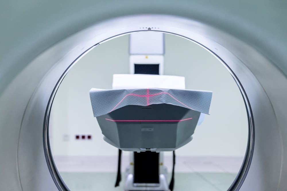
How is a CT scan performed?
Getting a CT scan is a painless and benign examination. While the patient is lying on their back in a tunnel, a ring rotates around the tunnel and delivers x-rays. Contrast can be given, usually by injection into the arm. The purpose of the injected iodine product is to promote the quality of the images.
When is the CT scan useful?
The CT scan can detect broken bones and brain damage, but it is also prescribed for infection, inflammation or exploration of the vessels. This test can also diagnose tumors and monitor their treatment, and research conditions such as Alzheimer’s disease and multiple sclerosis.
Risks and contraindications linked to the CT scan
Due to the exposure to x-rays, a CT scan is contraindicated for pregnant women. If iodine is injected, it can cause allergic reactions, although this is quite rare. Preventive treatment can be prescribed the day before the exam.
MRI: what is it?
The MRI is an extremely precise examination of your health. Unlike a CT scan, an MRI has the advantage of not exposing the patient to x-rays and MRIs are more detailed in their images.
What is an MRI used for?
An MRI, for magnetic resonance imaging, allows a detailed analysis of tissues such as muscles, heart, spinal cord, brain or tendons. This is why MRI is used for the exploration of many vascular, gynecological, gastric, cerebral and even joint diseases.
How does an MRI work?
The patient is laid down in a tube, similar to a CT scan. The medical team, present behind glass in an examination room, can observe the patient and communicate with him via a microphone system. If the information provided by the MRI is sufficient, no injection is necessary.
But in some cases, MRI may require injection of a gadolinium-based contrast medium. This is the case when it is necessary to obtain more details about an anomaly. The duration of an MRI varies between 10 and 20 minutes compared to only one to four minutes for a CT scan. MRI is not recommended for claustrophobic people and patients with a pace maker or certain models of heart valves.
What technology is used?
MRI is based on the property of the nuclei of hydrogen atoms in the human body to emit detectable signals when placed in a strong magnetic field and subjected to a radio frequency pulse capable of making them resonate.
These signals emitted by the hydrogen protons are then converted into images using a powerful computer system. The advantage of MRI is that it does not use x-rays, so it is harmless to health and is completely painless.
Contraindication differences between a CT scan and an MRI
MRI: contraindications
Even though a magnetic field is harmless to the patient, there are some contraindications associated with MRI.
No material likely to be exposed to a magnetic field can in fact enter the device’s magnetic field: this presupposes having suitable equipment (infusor, respirator, etc.) explaining that the scanner is often used as a first-line treatment. during an emergency.
For the patient, the presence of ferro-metallic bodies capable of moving with the MRI constitutes a formal contraindication: cerebral vascular clips (cerebral aneurysm), wearing of a cardiac pacemaker (Pace Maker), a neurosimulator , a cochlear implant, metal implants, intraocular foreign bodies.
Many implants (dental implants for example) are not metallic and therefore do not constitute an absolute contraindication: in all cases, the radiologist studies the medical file and the nature of the material implanted in the patient, to verify the absence of contraindications.
CT Scanner: contraindications
As X-rays present teratogenic risks for the fetus, the scanner may be contraindicated in pregnant women, depending on the irradiated area and the stage of pregnancy: an experienced radiologist is in perfect control of these contraindications.
There may be other contraindications related to the injection of the contrast product (myeloma, insulin treatment, allergies, breastfeeding, etc.).
Through his expertise, the radiologist determines whether this is an absolute and relative contraindication.
CT or MRI therefore remain two different medical imaging examinations, the indications for which are determined by the attending physician and the radiologist.
Difference Between CT Scan and MRI: Imaging Techniques and Applications
Medical imaging plays a crucial role in diagnosing and monitoring various health conditions, and two commonly used modalities are Computed Tomography (CT) scans and Magnetic Resonance Imaging (MRI). Each technique offers unique advantages and is best suited for specific imaging needs.
CT Scan (Computed Tomography): CT scans use X-ray technology to create detailed cross-sectional images of the body. They are particularly well-suited for visualizing dense structures like bones and are commonly used to examine the following:
Head and Brain: CT scans can identify intracranial bleeding, tumors, and structural abnormalities in the brain.
Chest: They are useful for diagnosing lung conditions, identifying lung nodules, and assessing the heart’s blood vessels.
Abdomen and Pelvis: CT scans help visualize the organs in the abdominal cavity, detect abdominal masses or tumors, and evaluate the gastrointestinal tract.
Spine: They provide detailed images of the spine’s vertebrae, discs, and surrounding soft tissues, aiding in diagnosing spinal fractures and herniated discs.
MRI (Magnetic Resonance Imaging): MRI uses strong magnetic fields and radio waves to produce detailed images of soft tissues and organs. It is particularly effective in imaging the following areas:
Brain and Nervous System: MRI can visualize the brain, spinal cord, and nerves, making it valuable for diagnosing multiple sclerosis, stroke, and brain tumors.
Muscles and Joints: It provides detailed images of muscles, tendons, ligaments, and joints, aiding in the assessment of sports injuries and musculoskeletal conditions.
Abdominal Organs: MRI is used to examine the liver, kidneys, pancreas, and reproductive organs, helping detect tumors, cysts, and other abnormalities.
Pelvic Organs: It is commonly used to image the uterus, ovaries, prostate, and bladder, assisting in diagnosing conditions like fibroids and prostate cancer.
Choosing Between CT Scan and MRI: The choice between CT scan and MRI depends on the specific clinical question, the part of the body being imaged, and patient considerations. CT scans are well-suited for bone imaging and emergencies due to their speed, while MRI excels in imaging soft tissues and is preferred when avoiding ionizing radiation is important.
Ultimately, healthcare providers determine the appropriate imaging modality based on the patient’s medical history, symptoms, and the information needed for an accurate diagnosis and treatment plan. Both CT scans and MRI play essential roles in modern medical practice, contributing to improved patient care and outcomes.
Information: Cleverly Smart is not a substitute for a doctor. Always consult a doctor to treat your health condition.
Sources: PinterPandai, Oesophageal Patients Association (OPA), Healthline, Memorial Sloan Kettering Cancer Center (MSKCC)