Hemianopsia
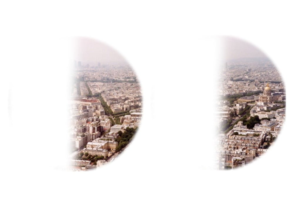
Hemianopsia is decrease or loss of vision in half of the visual field in one or both eyes.
Depending on the case, it can be considered unilateral or bilateral, lateral or altitudinal, homonymous or heteronymous. This vision disorder is caused by nerve damage to the optic nerve.
Among the branches of semiology, is clinical semiology, which refers to the study of the signs and symptoms through which a disease manifests itself. In the case of ophthalmology, it is very useful to know at least the most general and simple techniques of examining a patient.
Definition: what is hemianopsia?
Hemianopia is a vision disorder that involves a visual hemfield, which is only one half of the visual field of the eye. It can be unilateral, affecting vision in one eye, or bilateral, affecting visual acuity in both eyes.
In some cases, hemianopsia can be distinguished from quadranopsia. The latter concerns only part of the visual hemisphere. Depending on its location, the quadranopsy can be said to be left or right, lower or upper.
Symptoms
- Missing parts of words or parts of an eye chart on the side when reading.
- Lost part of the picture when watching TV.
- Sensation of problem with only one eye.
- Loss of half of the visual field.
- Frequent turning of the head to the side where hemianopia is present.
- Collisions with objects: affected people do not see obstacles or bump into road signs on the sidewalk
- Inability to find items: patients do not immediately find items on a shelf or table and have to search for them
reading and writing difficulties: the inability to read profoundly affects the quality of life.
It is therefore important to train patients with hemianopsia in reading and writing.
Caused
How is hemianopsia manifested?
Hemianopia is caused by nerve damage to the optic nerve, the sensory cranial nerve connecting each eyeball to the brain. Depending on the area of the affected nerve, this visual disturbance can manifest itself differently in the eye. The visual field of the eye can be divided into:
- a vertical axis, with a lower visual hemisphere and an upper hemisphere;
- a horizontal axis, with a left visual hemfield and a right visual hemfield, or with an exterior visual hemfield (temporal visual hemfield located near the temples) or interior (nasal visual hemisphere).
What are the different types of hemianopsia?
Depending on the case, it is possible to distinguish several types of hemianopsia:
- homonymous lateral hemianopia, the most common, when the same visual hemfield, either left or right, is affected in both eyes;
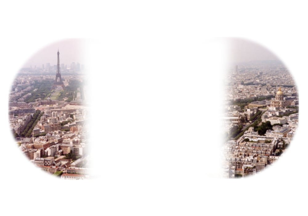
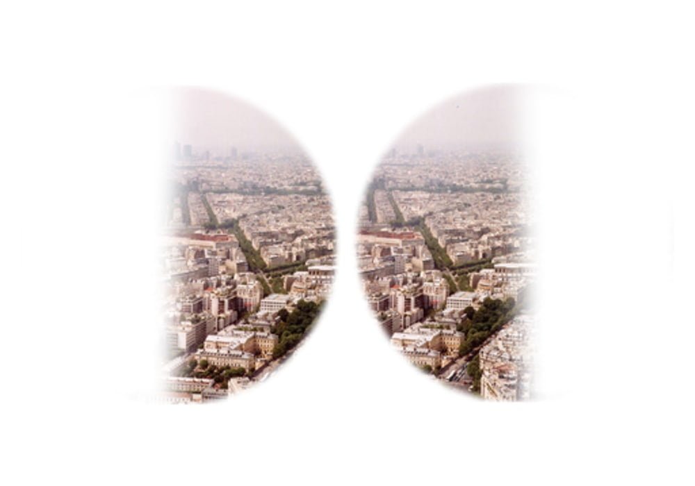
- heteronymous lateral hemianopia, more often called heteronymous hemianopia, when vision in both eyes is impaired either in the temporal visual hemianopia (bilateral hemianopia) or in the nasal visual hemianopia (binasal hemianopia);
- If occurs in both eyes it is called bilateral hemianopsia.
- altitudinal hemianopia, a rare form, when the upper or lower visual hemfield is affected.
Subtypes include:
- altitudinal hemianopsia, characterized by a visual defect above or below the horizontal meridian of the visual field;
- homonymous hemianopsia refers to a visual defect that affects both eyes equally, and occurs either to the left or right of the midline of the visual field;
- binasal hemianopsia consists of loss of vision in the nasal hemifields of both eyes;
- bitemporal hemianopsia is the bilateral loss of vision of the temporal fields;
- quadrantanopsia refers to loss of vision in one-fourth of the visual field in one or both eyes.


Hemianopsia: diagnosis
When a person has symptoms that suggest they are suffering from hemianopsia, the ophthalmologist will seek to examine the extent of their visual field.
For this, he/she can use a visual test consisting in asking the patient to fix a target placed in front of him, then to send light signals to different places to detect possible blind areas.
Secondly, magnetic resonance imaging (MRI) will be considered to detect the existence of a brain lesion that could explain the hemianopsia.
VISUAL FIELD INSPECTION
Visual field examination was carried out in a dark room.
Individuals taking this exam are first asked to perform a dark adaptation for 20 minutes.
Visual field studies are based on:
Campimetry: exploration of the mid-zone of the visual field.
This is done with a campimeter and basically makes it possible to highlight the central and paracentral (around the center) scotoma (gap in the visual field).
Perimetry: exploration of the entire visual field.
This is done using a perimeter (an optical instrument used in ophthalmology to explore the breadth of the visual field). Several types of perimeter are available to ophthalmologists (ophthalmologists):
The Goldmann machine, which has a small dome with the subject staring intently in the center and where the light points move at different sizes and with varying light intensity
Landolt Visual Acuity Scale: The Landolt Visual Acuity Scale, used to check visual acuity, consists of a series of optotypes that are all identical (broken circles, called Landolt rings) but whose orientations vary. It was developed in 1888 by Edmond Landolt (1846-1926), a Swiss ophthalmologist. It is a vision test which is useful especially for children and illiterate. In practice, this is the most appropriate measurement of visual acuity because it removes the effects of memorization and interpretation, unlike letters and numbers.
Perimeter Boundary Magitot: from the scope of Ferree and Rand, consists of: a perimeter arc with a radius of 33 cm, painted in a neutral gray color, allowing the peripheral boundaries of the visual field to be defined as a perimeter arc of the same radius; it is none other than the center of the hemisphere where the arc is, reserved as full screen up to 30 degrees; it is used for scotometer.
Explanation: what are the causes of hemianopsies?
The nerve damage that causes hemianopsia can have several causes. These are generally of tumor, vascular or traumatic origin.
1. Causes of tumor origin
The development of a tumor in the brain can compress the optic nerves and cause homonymous or heteronymous lateral hemianopsia. Visual loss can, for example, be due to the development:
a pituitary adenoma, a benign, non-cancerous tumor of the pituitary gland, an endocrine gland in the brain;
a meningioma, a usually benign tumor that develops in the meninges, membranes covering the brain and spinal cord;
glioma, a benign or malignant tumor that appears in the glial tissue, the tissue that supports neurons.
2. Causes of vascular origin
Lateral hemianopia of the same name can also be the result of a stroke. A stroke is caused by a blood flow disorder in the brain.
3. Causes of traumatic origin
In some cases, the nerve damage that causes hemianopsia is due to head trauma.
Evolution: what is the risk of complications?
The consequences and course of hemianopsia depend on many parameters, including the origin of the nerve damage. In more severe cases, the visual disturbance may worsen and become irreversible. Early medical treatment is necessary to limit the risk of complications.
Treatment: what are the solutions in case of hemianopia?
Treatment for hemianopsia varies depending on the underlying disease. If this disorder develops, it should be considered a symptom of another disease and not of the disease itself. It is recommended that you see a specialist so that they can do an eye exam.
The medical management of hemianopsia is usually based on the treatment of the nerve damage. Depending on its origin, it may sometimes require surgery.
Treatment of hemianopia is usually accompanied by optical rehabilitation sessions. Carried out by an orthoptist, this rehabilitation allows the patient to adapt to his visual field.
Treat brain damage
Therapeutic treatment related to the associated brain injury may also contribute to vision changes and symptoms of hemianopia. For example, the removal of a brain tumor or the surgical and medical treatment of brain inflammation can help improve associated visual deficits.
Eye Diseases and Common Eye Problems (List of eye diseases and disorders)
Information: Cleverly Smart is not a substitute for a doctor. Always consult a doctor to treat your health condition.
Sources: PinterPandai, Cleveland Clinic, Radiopaedia, University of Utah Health Care