What are retinal diseases?
The retina, which lines the back of the eye, contains nerve cells that receive light. They translate it into electrical signals that travel to the brain via the optic nerve. There are many types of retinal diseases.
When the cells degenerate or no longer function, blind areas of the visual field appear.
Many pathologies can affect this area of the eye: AMD, diabetic retinopathy, retinitis pigmentosa…
Different types of retinal diseases
Macular degeneration
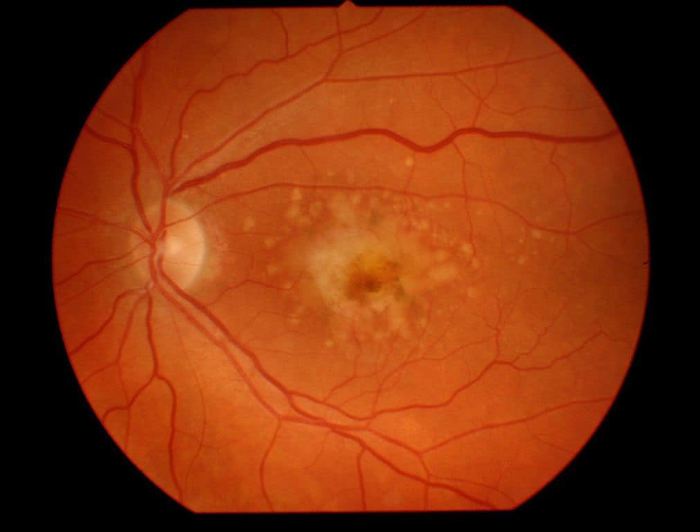
In this pathology, the decline in visual acuity is due to damage to the macula, the central area of the retina which transmits most of the visual information to the brain. At an advanced stage, a blind spot appears in the center of the field of vision. Peripheral vision is preserved. The disease comes in two forms:
the so-called “dry” or atrophic form is the most frequent (two thirds of cases). Slowly evolving, it is the consequence of the progressive disappearance of the cells of the macula.
the “wet” or exudative form, observed in a third of cases, progresses rapidly. It is caused by the formation of abnormal blood vessels under the retina.
The risk factors identified are age, genetic predisposition, smoking and probably excessive sun exposure during youth.
Diabetic retinopathy
This complication of diabetes is linked to excessive sugar concentration in the small blood vessels of the retina, which leads to their degradation. The lack of oxygen supply induces the formation of new, more fragile blood vessels. Their rupture and the ensuing micro-haemorrhages can lead to true retinal detachment.
Another characteristic of these new vessels: they are more permeable and tend to “leak”. The resulting accumulation of blood and fluids causes edema resulting in a sharp drop in visual acuity.
Screening for diabetic retinopathy requires close monitoring. Diagnosed in time, it is controlled in most cases without progressing to loss of vision.
Hereditary diseases of the retina
Retinitis pigmentosa, Leber congenital amaurosis or Stargardt disease are rare genetic diseases that also affect the retina.
Retinitis pigmentosa is one of the most common, with one in 3,500 people affected. It includes several pathologies that initially result in a decrease in vision during a drop in light. They then progress to a decrease in peripheral vision during the day and gradually lead to blindness.
Read also: Eye Diseases and Common Eye Problems (List of eye diseases and disorders)
Leber’s congenital amaurosis is one of the main causes of blindness in children (it is found in 10 to 20% of blind children). For the moment it is incurable, and has been the subject of increased research in recent years. Same observation for Stargardt’s disease, for which the management methods are currently palliative.
Retinitis
Retinitis is inflammation of the retina. The course of the disease can develop differently and ranges from almost no impairment of the patient to complete blindness due to degeneration of the retina. Retinitis is often accompanied by inflammation of the choroid.
Chorioretinitis | Inflammation of the eye behind the retina
Chorioretinitis is the medical term for inflammation of the retina (→ retinitis ) and the choroid . Rarely is the term used upside down as retinochoroiditis. The course of the disease can develop differently and ranges from almost no impairment of the patient to complete blindness due to degeneration of the retina.
Cytomegalovirus retinitis
The disease is often triggered by general infections, such as toxoplasmosis or tuberculosis and late syphilis. Bacterial-metastatic inflammations are less common. In AIDS patients or immunocompromised people, chorioretinitis is often caused by the human cytomegalovirus.
They are rarely caused by widespread bacterial infections. AIDS or immunosuppressed patients often have chorioretinitis caused by cytomegalovirus . It can also be caused by the Arenavirus that causes lymphocytic choriomeningitis, transmitted by secretions from rats and mice.
Retinitis pigmentosa
The term retinopathy pigmentosa or retinitis pigmentosa (RP) describes a retinal degeneration caused by inheritance or spontaneous mutation , in which the photoreceptors are destroyed. One speaks of pseudoretinitis pigmentosa (or phenocopy) when non-hereditary diseases show symptoms of retinopathy pigmentosa, for example due to toxicity (e.g. due to thioridazine , chloroquine).
Central serous chorioretinopathy
Central serous chorioretinopathy is fluid accumulated in the subretinal space. It is an eye disease that causes visual impairment, usually temporary, usually in one eye. Read also: Central Serous Chorioretinopathy (Retinal Disease)

Retinal tear
A retinal tear occurs when the clear, gel-like substance in the center of your eye (vitreous) shrinks and tugs on the thin layer of tissue lining the back of your eye (retina) with enough traction to cause a break in the tissue. It’s often accompanied by the sudden onset of symptoms such as floaters and flashing lights.
Retinal detachment
A retinal detachment is defined by the presence of fluid under the retina. This usually occurs when fluid passes through a retinal tear, causing the retina to lift away from the underlying tissue layers. Initial detachment may be localized, but without rapid treatment the entire retina may detach, leading to vision loss and blindness. It is a surgical emergency.
Retinal cancer
The retina is a transparent and light-sensitive membrane at the back of the eye. Retinal cancer usually begins in the choroid, the dense vascular layer that nourishes the retina. The choroid is located between the retina and the sclera (the outer, white covering of the eye). Because retinal vasculature depends on the choroid, changes in the choroid by the cancer process can affect vision.
Epiretinal membrane
Epiretinal membrane is a delicate tissue-like scar or membrane that looks like crinkled cellophane lying on top of the retina. This membrane pulls up on the retina, which distorts your vision. Objects may appear blurred or crooked.
Retinitis pigmentosa
Retinitis pigmentosa is an inherited degenerative disease. It slowly affects the retina and causes loss of night and side vision.
Retinoschisis
Retinoschisis is a splitting of the retina into two layers with layer ruptures and mostly mutual retinal defects (perforated, vascular membrane formations). It is characterized by degeneration linked to a mutation of the retina gene. The major consequence of this disease is retinal detachment and a decrease in visual acuity.
Ocular ischemic syndrome
Central retinal artery obstruction is a serious eye disease caused by the closure of this artery to blood flow. The main symptom is an abrupt onset of vision loss in the affected eye.
Central retinal vein occlusion
Central retinal vein occlusion, also CRVO, is when the central retinal vein becomes occluded, usually through thrombosis. The central retinal vein is the venous equivalent of the central retinal artery and both may become occluded. Since the central retinal artery and vein are the sole source of blood supply and drainage for the retina, such occlusion can lead to severe damage to the retina and blindness, due to ischemia (restriction in blood supply) and edema (swelling).
CRVO can cause ocular ischemic syndrome. Nonischemic CRVO is the milder form of the disease. It may progress to the more severe ischemic type. CRVO can also cause glaucoma.
Central retinal artery occlusion
Central retinal artery occlusion is a a rare and severe disease of the eye where the flow of blood through the central retinal artery is blocked (occluded). There are several different causes of this occlusion; the most common is carotid artery atherosclerosis.
Branch retinal artery occlusion
Is a rare retinal vessel disorder in which a branch of the central retinal artery becomes occluded. The typical symptom is a sudden, painless loss of vision in the visual field covered by the blocked artery. Patients can usually determine the exact time and extent of vision loss.
The most prominent medical result is the bleaching of the retina in the area corresponding to the area that suffers from ischemia . Retinal bleaching disappears in the chronic stage.
Retinopathy
A retinopathy (from πάθος, pathos, “suffering” and Latin retina retina ) is damage to the retina of the eye.
A distinction is made according to the cause
- Diabetic retinopathy (retinal disease caused by diabetes mellitus )
- Retinopathia centralis serosa (swelling-related retinal detachment)
- Retinopathia pigmentosa (hereditary disease of the retina)
- Hypertensive retinopathy (retinal disease caused by high blood pressure)
- Eclamptic retinopathy (retinal disease that occurs in pregnant women as part of eclampsia )
- Retinopathia praematurorum (retinal disease in premature babies)
- Retinopathia solaris or photic retinopathy (retinal damage caused by sunlight or laser)
- Purtscher’s retinopathy is a disease where part of the eye (retina) is damaged. Usually associated with severe head injuries, it may also occur with other types of trauma, such as long bone fractures, or with several non-traumatic systemic diseases.
- Retinopathy of prematurity (ROP), also called retrolental fibroplasia (RLF) and Terry syndrome, is a disease of the eye affecting prematurely born babies generally having received neonatal intensive care, in which oxygen therapy is used due to the premature development of their lungs. It is thought to be caused by disorganized growth of retinal blood vessels which may result in scarring and retinal detachment. ROP can be mild and may resolve spontaneously, but it may lead to blindness in serious cases. Thus, all preterm babies are at risk for ROP, and very low birth-weight is an additional risk factor. Both oxygen toxicity and relative hypoxia can contribute to the development of ROP.
- Bietti’s crystal dystrophy is a very rare congenital disease of the retina with tapetoretinal degeneration beginning in the third decade of life . The main symptoms are crystalline deposits of unclear composition in the rear parts of the retina, in the area of the corneal limbus and in the leukocytes . Next comes a sclerosis of the choroid vessels.
- Coats’ disease, is a rare congenital, nonhereditary eye disorder, causing full or partial blindness, characterized by abnormal development of blood vessels behind the retina. Coats’ disease can also fall under glaucoma. It can have a similar presentation to that of retinoblastoma (rare form of cancer that rapidly develops from the immature cells of a retina, the light-detecting tissue of the eye. It is the most common primary malignant intraocular cancer in children, and it is almost exclusively found in young children).
- Sickle cell retinopathy can be defined as retinal changes due to blood vessel damage in the eye of a person with a background of sickle cell disease. It can likely progress to loss of vision in late stages due to vitreous hemorrhage or retinal detachment. Sickle cell disease is a structural red blood cell disorder leading to consequences in multiple systems. It is characterized by chronic red blood cell destruction, vascular injury, and tissue ischemia causing damage to the brain, eyes, heart, lungs, kidneys, spleen, and musculoskeletal system.
Retinal haemorrhage
Is a disorder of the eye in which bleeding occurs in the retina, the light sensitive tissue, located on the back wall of the eye. There are photoreceptor cells in the retina called rods and cones, which transduce light energy into nerve signals that can be processed by the brain to form visual images.[2] Retinal hemorrhage can affect adults, and newborn babies and infants may also suffer from this disorder.
A retinal hemorrhage can be caused by several medical conditions such as hypertension, retinal vein occlusion (a blockage of a retinal vein), anemia, leukemia or diabetes.
Central serous retinopathy
Central serous chorioretinopathy is fluid accumulated in the subretinal space. It is an eye disease that causes visual impairment, usually temporary, usually in one eye. When the disorder is active it is characterized by leakage of fluid under the retina that has a propensity to accumulate under the central macula.
This results in blurred or distorted vision (metamorphopsia). A blurred or gray spot in the central visual field is common when the retina is detached. Reduced visual acuity may persist after the fluid has disappeared.
Macular edema
Is a collection of extracellular fluid (edema) in the area of the yellow spot (macula lutea) of the human eye. The reversible change is expressed by blurring of the field of vision in the zone of sharpest vision. The macula is a small area of the retina measuring approximately 1 mm very rich in visual cells, which is responsible for precision vision. When it is very close to the center of the retina and threatens visual function, it is called clinically significant macular edema. If left untreated, it can lead to permanent severe central visual acuity loss. Read also: Macular Edema | Eye Disorders | Symptoms, Causes, Treatment, Prevention
Epiretinal membrane or cellophane maculopathy (Macular pucker)
Is a pathology of the eye which consists in the development of a thin translucent membrane over the macula (the central area of the retina essential for vision). When this membrane contracts and curls, it causes a deformation and progressive distortion of the macula itself.
At the onset of the disease there is a slight distortion of the images and, above all, of the texts written with smaller characters (the lines will appear wavy). When the traction exerted by the membrane on the macula increases, we realize that reading the texts becomes more and more difficult. Eventually, the evolution of the disease leads to the vision of a spot that prevents central vision.
The diagnosis is made thanks to the examination of the fundus, which allows to visualize the membrane. However, confirmation must be obtained through OCT (Optical Coherence Tomography), an examination with which the extent of traction is assessed: it helps to control the evolution of the pathology over time.
Vitelliform macular dystrophy
Vitelliform macular dystrophy, is an irregular autosomal dominant eye disorder which can cause progressive vision loss. This disorder affects the retina, specifically cells in a small area near the center of the retina called the macula. The macula is responsible for sharp central vision, which is needed for detailed tasks such as reading, driving, and recognizing faces. The condition is characterized by yellow (or orange), slightly elevated, round structures similar to the yolk (Latin vitellus) of an egg.
Leber’s congenital amaurosis
Is a serious genetic disease of the retina described for the first time in 1869 by Théodor Leber which he called retinitis pigmentosa in utero.
Those affected are already significantly visually impaired or blindto the world, and the probability of subsequent siblings being affected is about 25%. More than 10% of all congenital blindness cases can be attributed to Leber’s congenital amaurosis.
Birdshot chorioretinopathy
Is a rare form of bilateral posterior uveitis affecting both eyes. It causes severe, progressive inflammation of both the choroid and retina.
Symptoms of this disorder include mottling (floating over vision), blurred vision, photopsia (flashes of light in the eyes), loss of color vision, and night blindness (inability to see in low-light conditions). Color points are seen on the retina in eye tests. Possible complete loss of visual acuity.
The disease’s name is given by the small colored dots on the retina, scattered in a pattern like birdshot, but these dots may not be present in early stages.
Birdshot retinochoroidopathy could show resistance to treatment. With immunosuppression therapy with oral corticosteroids, it could somehow be effective in slowing down the inflammatory process associated with the disease, preserving visual integrity as much as possible. Long-term use of such medications should be closely monitored, however, due to discomfort, and potentially debilitating and life-threatening side effects, subsequent birdshot retinochoroidopathy patients treated with immunomodulatory systemic corticosteroid therapy were treated. with systemic immunosuppressants.
Immunosuppressive drugs such as monoclonal antibodies, daclizumab, cyclosporine, and methotrexate have been tested as effective treatment options for shotgun retinochoroidopathy. Substantial reduction and even stabilization of vitreous inflammation and retinal vasculitis has been evidenced by electroretinography during daclizumab (IL-2 receptor blocker) therapy. This is also supported by the observation of elevated levels of IL-2 in the eyes of patients.
Information: Cleverly Smart is not a substitute for a doctor. Always consult a doctor to treat your health condition.
Sources: PinterPandai, Mayo Clinic, MedlinePlus, The American Society of Retina Specialists, Optometrists
Main photo source: Tmhlee / Wikimedia Commons (CC BY-SA 3.0)
Main photo description: this image shows the fundus of the eye (examination of the eye) viewed through a retinal camera, with signs of macular degeneration.