What is macular edema?
The macula is the central area of the retina or fundus and is the point where visual acuity is at its peak. An intact macula allows clear central vision, distinguishing details, reading and recognizing, for example, people’s faces. Macular edema is an increase in the volume of the macula, the central area of the retina, which is responsible for visual acuity. Visual symptoms cripple the lives of millions of patients with macular edema from chronic and sometimes acute retinal disease.
Macular edema is a frequent pathology and consists of an abnormal accumulation of fluid or “flooding” in the area of the macula due to an alteration in the permeability of the blood vessels that feed it.
Symptoms
It is usually painless and is characterized by blurred central vision, image distortion or waviness, color alteration (discolored or different vision) and is accompanied by difficulty in reading.
Causes
There are different eye pathologies that can cause macular edema, including: diabetic retinopathy (the most common), venous thrombosis or occlusion, uveitis (intraocular inflammation), following eye surgery, retinal dystrophies such as as retinitis pigmentosa, age-related macular degeneration, intraocular tumors, traction phenomena such as the epiretinal membrane (tissue that grows on the surface of the macula and alters its normal morphology) or even certain topical eye drops for the treatment glaucoma (prostaglandin analogues).
Cystoid Macular Edema (CME)
In some cases, this condition can occur after eye surgery. When macular edema appears after cataract surgery, it is called Cystoid Macular Edema (CME).
Although this visual condition can also be associated with other eye diseases, such as:
- Diabetic retinopathy
- Age-Associated Macular Degeneration
- Uveitis
- Certain types of retinitis
- Certain type of tumors in the eyes
- Genetic disorders, such as some forms of macular dystrophy
- This disorder can also be caused by eye injuries or the side effects of certain medications.
For people with diabetes, macular edema is usually the most common cause of vision loss.
Treatments
There are different options for treating macular edema, depending on the patient, the cause of the edema, as well as its degree of severity. The best treatment will be a personalized treatment for each case.
In some cases, the use of anti-inflammatory eye drops may be sufficient. Another form of treatment consists of injections of drugs both in the periocular area (around the eyeball) and intraocularly (injection of the drug into the eye).
These drugs, which may be corticosteroids or anti-angiogenics, act locally on the macula to reduce inflammation and fluid extravasation.
Laser photocoagulation is also useful for treating some cases of macular edema by “sealing” fluid leak points. Finally, in some cases, it may be necessary to perform retinal and vitreous surgery to treat macular edema caused by macular traction, in which a tissue present on the surface of the retina is responsible for the alteration of the macula.
Treatments for Cystoid Macular Edema (CME)
The cause should be treated when possible (diabetes, uveitis) and/or associated treatment with acetazolamide (Diamox®), and local non-steroidal or steroidal anti-inflammatory drugs.
Prevention
The different prevention strategies will depend on the cause of the macular edema.
People with diabetes must ensure good metabolic and glycemic control, and take into account that this complication can appear at any stage of diabetic retinopathy and not only at an advanced stage.
It is often associated with vascular pathologies, it is also recommended to adopt healthy lifestyle habits (healthy diet, regular exercise, etc.) that avoid predisposing factors such as high blood pressure and excess fat. cholesterol.
Likewise, to avoid irreversible deterioration of the macula, it is important to carry out regular ophthalmological checks allowing a complete examination of the fundus and its blood vessels to detect any abnormality as soon as possible. In this sense, an annual examination is recommended despite the absence of symptoms.
Macular edema, in some cases, is associated with other pathologies such as diabetes. In these cases, it will be necessary to control this disease by ensuring that the sugar level is correct.
It cannot be prevented, although it can be detected quickly if periodic ophthalmological checks are carried out, especially in the presence of predisposing factors.
Sources: PinterPandai, National Eye Institute, American Academy of Ophthalmology, The American Society of Retina Specialists, Retina UK
Main photo source: Community Eye Health / Flickr (CC BY-NC 2.0)
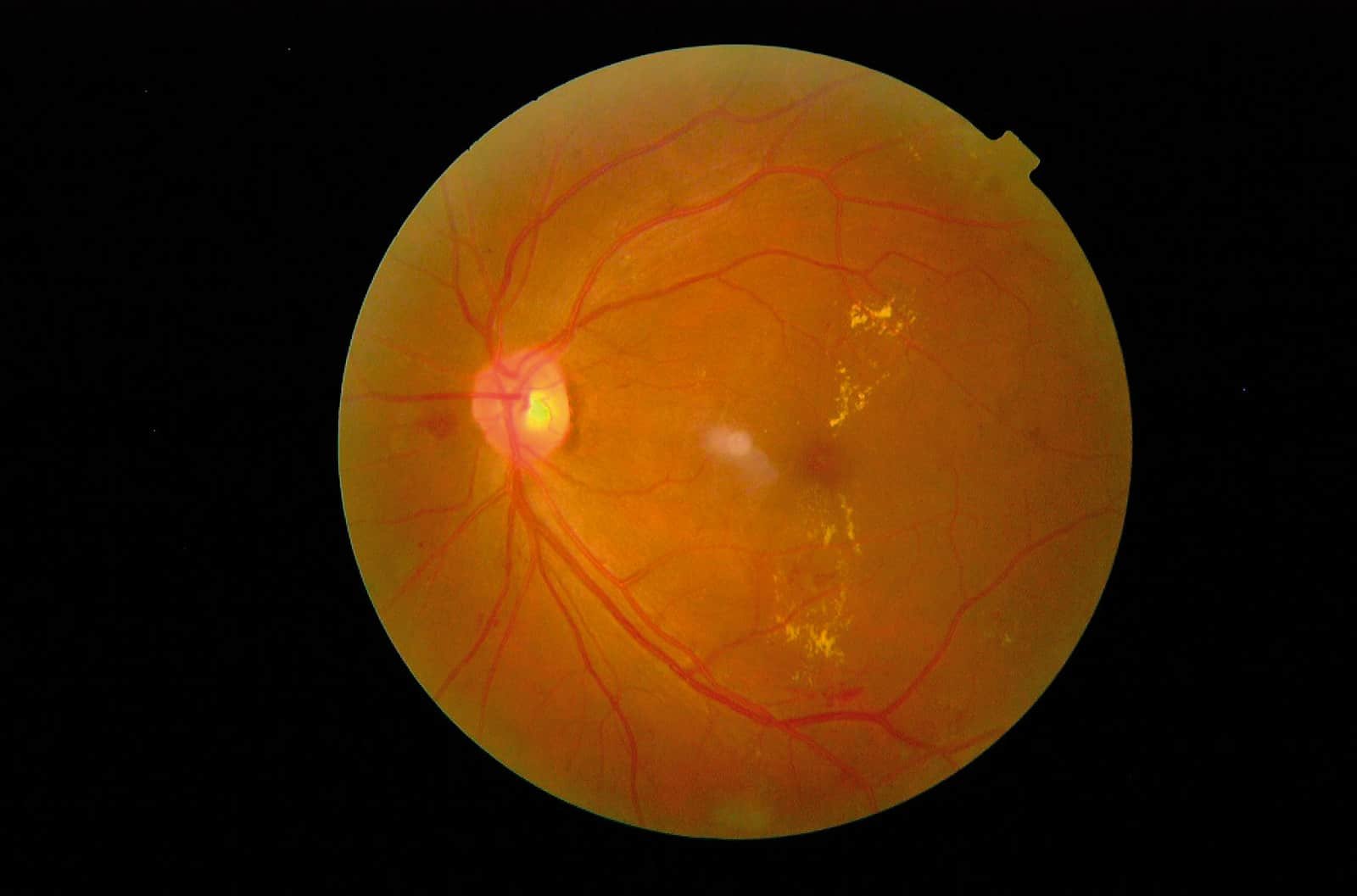
Title: Clinically significant macular oedema with hard exudates encroaching on the fovea. Photo: Sangita Sagar.
Main photo description: photo shows clinically significant macular oedema with hard exudates encroaching on the fovea, identified using photographic screening in an asymptomatic patient. Photo: Sangita Sagar. Published in: Community Eye Health Journal Vol. 21 No. 66 JUNE 2008 www.cehjournal.org