Teratoma
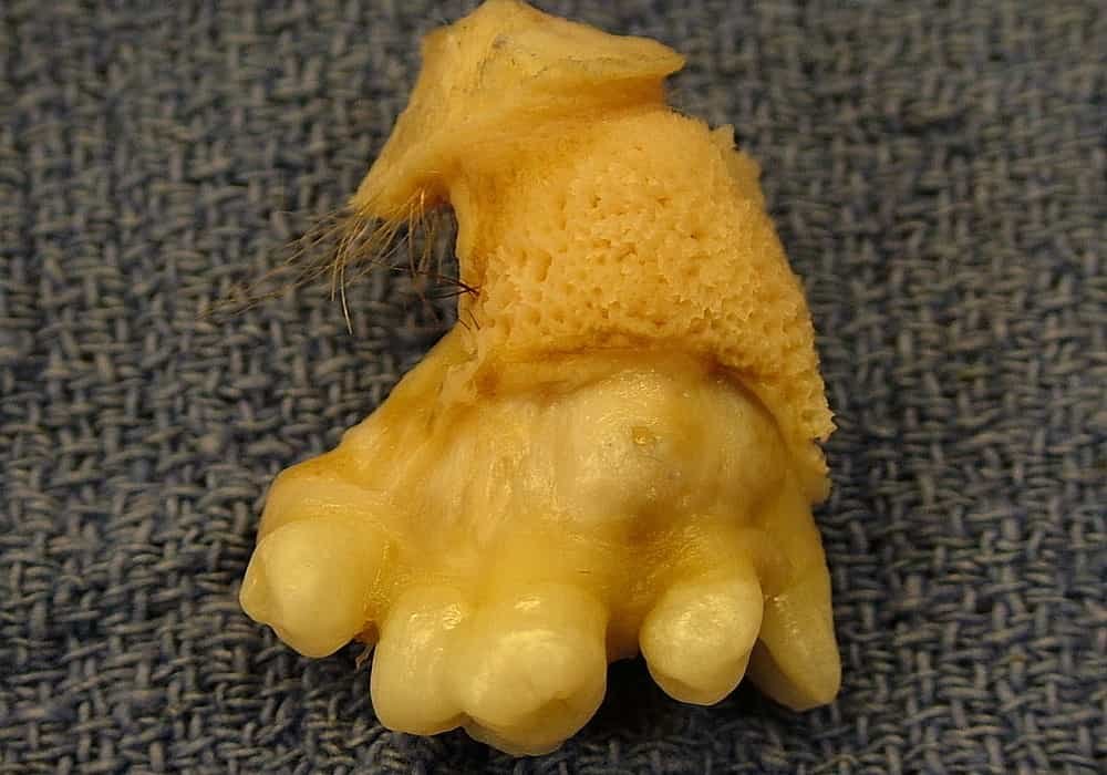
Teratoma is a tumor that can be benign or malignant (cancerous) and can contain different types of tissue, such as hair, muscle and bone or teeth.
These tumors are said to be germinal because they develop from primordial germ cells (cells that produce gametes: spermatozoa in men and ova in women).
They often seem to be associated with glandular hormonal activity (eg pineal gland).
The two most common forms are:
- ovarian teratoma in women;
- testicular teratoma in men.
However, teratomas can also develop in other areas of the body. We can in particular distinguish:
- sacrococcygeal teratoma (between the lumbar vertebrae and the coccyx);
- cerebral teratoma, which mainly occurs in the epiphysis (pineal gland);
- mediastinal teratoma, or teratoma of the mediastinum (region of the chest located between the two lungs).

Read also:
- Testicular Cancer | Symptoms, Stages, Types, Diagnoses, Chances of Surviving, Treatments
- Ovarian Cancer | Symptoms, Stages, Types, Diagnoses, Chances of Surviving, Treatments
- Brain Cancer | Symptoms, Stages, Types, Diagnoses, Chances of Surviving, Treatments
Classification of teratomas
Teratomas can be very different. Some are benign while others are malignant (cancerous).
Three types of teratomas are defined:
- mature teratomas which are benign tumors made up of well-differentiated tissue;
- immature teratomas which are malignant tumors (cancer) made up of immature tissue still resembling embryonic tissue;
- monodermal or specialized teratomas which are rare forms which may be benign or malignant.
Teratomas with malignant (cancer) transformation
In rare cases, mature or immature teratomas can undergo malignant transformation. Teratomas with a malignant transformation in the conventional sense represent a well-circumscribed unit whose name refers to the malignant transformation of a somatic teratomatous component within a non- seminomatous germ cell tumor into a histology indistinguishable from a somatic malignancy. Examples of these histologically transformed cell types include
- Rhabdomyosarcoma (RMS)
- Primitive neuroectodermal tumors (PNET): develop from immature nerve cells in the brain
- Enteric adenocarcinomas
- Leukemia
Cause of teratomas
They are characterized by the development of abnormal tissue. The origin of this abnormal development has not yet been established.
People affected by teratomas
It represent 2 to 4% of tumors in children and young adults. They represent 5 to 10% of testicular tumors. In women, mature cystic teratomas account for 20% of ovarian tumors in adults and 50% of ovarian tumors in children. Brain teratoma accounts for 1 to 2% of brain tumors and 11% of childhood tumors. Diagnosed before birth, sacrococcygeal teratoma can affect up to 1 in 35,000 newborns.
Diagnosis of teratomas
The diagnosis is usually based on medical imaging (X-ray, ultrasound, bone scans, MRI, and CT). However, exceptions exist depending on the location of the teratoma and its development. Blood tests for tumor markers can, for example, be carried out in certain cases. Biopsy to see if the teratoma is cancerous.

Symptoms of teratomas
Some of them may go unnoticed while others will cause great discomfort. Their symptoms depend not only on their form but also on their type.
Possible swelling
Some teratomas can manifest as swelling of the affected area. For example, an increase in testicular volume can be observed in testicular teratoma.
Other associated signs
In addition to the possible swelling in certain locations, a teratoma can induce other symptoms such as:
- abdominal pain in ovarian teratoma;
- respiratory discomfort when the teratoma is localized in the mediastinum;
- urinary disorders or constipation when the teratoma is localized in the region of the coccyx;
- headache, vomiting and visual disturbances when the teratoma is located in the brain.
Risk of complications
The presence of a teratoma may pose a risk of complications. In women, ovarian teratoma can lead to several complications such as:
- an adnexal torsion which corresponds to a rotation of the ovary and the fallopian tube;
- infection of the cyst;
- a ruptured cyst.
Treatments
The management of this disease is mainly surgical. The operation involves removing the teratoma. In some cases, surgery is supplemented by chemotherapy. This relies on chemicals to destroy diseased cells.
Prevention
The mechanisms involved in the development of a teratoma are not yet fully understood and that is why there is no specific prevention.
Other teratomas and their characteristics
Sacrococcygeal teratoma
Sacrococcygeal teratoma is a benign cystic teratoma of the fetus affecting the sacrococcygeal region (between the lumbar vertebrae and the coccyx):
It is diagnosed during fetal life, during an obstetric ultrasound.
It can be associated with spina bifida (incomplete development of the spine).
Sacrococcygeal teratoma affects 1 in 35,000 to 40,000 births, and 75% of affected fetuses are female.
It is most often a benign tumor, which develops around the pelvic region, either outside the body or inside the abdomen.
It can be very large (up to half the baby’s weight), causing compression of the abdominal organs (digestive tract, kidneys). Highly vascularized, it can cause circulatory disorders at birth and require surgery from birth.
The discovery of a sacrococcygeal teratoma during pregnancy leads to weekly ultrasound monitoring and a delivery usually scheduled by cesarean section.
The main complications for the fetus are not systematic, but they are serious:
- hydramnios (excess amniotic fluid);
- hydrops fetalis (generalized edema of the fetus);
- bleeding inside the teratoma;
- fetal death in utero.
The only possible treatment is surgery, performed soon after birth. Surgical reconstruction of the buttocks is often necessary after surgery to remove the tumor. The risk of recurrence is estimated at 10% on average.
Brain teratoma
Most often it develops in the epiphysis (or pineal gland), but it can also be located in the spinal cord, the base of the brain, or near the pituitary gland. Brain teratoma accounts for 1 to 2% of brain tumors but 11% of childhood tumors.
They are responsible for the following symptoms:
- intracranial hypertension;
- eye movement disorders (oculomotor disorders);
- endocrine disorders;
- sensory or motor disorders;
- psychiatric signs.
The diagnosis of cerebral teratoma is based on imaging (MRI, magnetic resonance imaging) and blood tests for tumor markers.
Treatment is based on surgery to remove the tumor, with a good prognosis (60 to 100% survival after 5 years). Headaches, eye movement disorders, or even a depressive syndrome may persist following surgery. Radiotherapy may be considered in addition to surgery.
Cervical teratoma
Cervical teratoma is a very rare tumor in newborns. Cervical teratoma is a mixed teratoma, associating mature and immature structures:
It is positioned either laterally at the level of the neck, or axially at the level of the pharynx.
It is most often detected during an obstetric ultrasound. An MRI (magnetic resonance imaging) and / or a CT scan can confirm the diagnosis.
Large tumors can cause compression of the airways (especially the trachea), which can cause respiratory distress in the newborn baby at birth.
Treatment is based on taking care of the child at birth (use of a caesarean section, management of potential respiratory distress) and surgery immediately after birth. Chemotherapy may accompany surgery if there is tumor residue after surgery.
Orbital teratoma
This orbital type is a rare tumor, mature or immature, affecting children from birth. Orbital teratoma causes:
- significant distension of the eyelids;
- exophthalmos (protrusion of the eye) or even disappearance of the eyeball;
- decreased visual acuity;
- oculomotor paralysis (inability to move the eyes).
The teratoma evolves very quickly, it can take the size of a tangerine, and be life threatening to the child. Treatment is based on surgery, which should be done as soon as possible after birth. The risk of recurrence is very low.
Mediastinal teratoma (this is an area in the middle of the chest that separates the lungs)
It can be benign, malignant, or mixed. Mediastinal malignant teratoma is a rare, highly progressive malignant tumor, affecting mainly young men. Benign mediastinal teratomas are either mature multi-tissue teratomas or single-tissue dermoid cysts.
Mediastinal teratoma causes compression of the organs located in the mediastinum, causing the symptoms that reveal the tumor:
- the lungs, the trachea and the two main bronchi;
- the heart ;
- the esophagus.
The diagnosis is made in the presence of calcifications on an x-ray. The only possible treatment is surgery to remove the tumor.
Only the analysis of the tumor after surgery can determine with certainty whether the tumor is benign or malignant.
Cardiac teratoma
This type is a tumor of complex structure, very rare, usually very serious, affecting children under 1 year of age. Cardiac teratoma can be located:
- most often in the pericardium, near the root of the aorta or pulmonary artery;
- more rarely, inside the heart near the wall of the atria or ventricles.
Cardiac teratoma is a large tumor (2 to 15 cm), usually immature. The diagnosis is often made by an obstetric ultrasound showing the tumor and sometimes an effusion of the pericardium (abnormal fluid infiltration).
The main risks are:
- in the fetus, foetoplacental hydrops (generalized edema of the fetus) or death in utero;
- in the newborn, respiratory distress, cyanosis and / or heart failure.
Treatment is based on surgery, which is particularly delicate for teratomas located inside the heart. After surgery, the prognosis is excellent for the child.
Retroperitoneal (area in the back of the abdomen behind the peritoneum (the tissue that lines the abdominal wall and covers most of the organs in the abdomen)
Retroperitoneal teratoma can be benign or malignant and can affect adults and very rarely, newborns and children, with a finding during obstetric ultrasounds:
These tumors grow within the peritoneum (in the abdomen).
The gradual increase in tumor size eventually causes compression of nearby organs.
In adults, the symptoms caused by this compression phenomenon are often the cause of the diagnosis. The diagnosis is confirmed by imaging tests (ultrasound, CT scan and MRI).
Surgical removal of the tumor is the only possible treatment and should be done before the tumor becomes too large.
Sources: PinterPandai, Healthline, Cancer.org